04 Januari, 2017

Understanding Cancer


What Is Cancer?

In the most basic terms, cancer refers to cells that grow out-of-control and invade other tissues. Cells may become cancerous due to the accumulation of defects, or mutations, in their DNA. Certain inherited genetic defects (for example, BRCA1 and BRCA2 mutations) and infections can increase the risk of cancer. Environmental factors (for example, air pollution) and poor lifestyle choices—such as smoking and heavy alcohol use—can also damage DNA and lead to cancer.
Most of the time, cells are able to detect and repair DNA damage. If a cell is severely damaged and cannot repair itself, it usually undergoes so-called programmed cell death or apoptosis. Cancer occurs when damaged cells grow, divide, and spread abnormally instead of self-destructing as they should.


Malignant Tumors Vs. Benign Tumors

A tumor is an abnormal mass of cells. Tumors can either be benign (non-cancerous) or malignant (cancerous).

>> Benign Tumors

Benign tumors grow locally and do not spread. As a result, benign tumors are not considered cancer. They can still be dangerous, especially if they press against vital organs like the brain.

>> Malignant Tumors

Malignant tumors have the ability to spread and invade other tissues. This process, known as metastasis, is a key feature of cancer. There are many different types of malignancy based on where a cancer tumor originates.


Cancer Metastasis

Metastasis is the process whereby cancer cells break free from a malignant tumor and travel to and invade other tissues in the body. Cancer cells metastasize to other sites via

the lymphatic system and the bloodstream. Cancer cells from the original—or primary—tumor can travel to other sites such as the lungs, bones, liver, brain, and other areas. These metastatic tumors are "secondary cancers" because they arise from the primary tumor.

Metastatic cancer retains the name of the primary cancer. For example, bladder cancer that metastasizes to the liver is not liver cancer. It is called metastatic bladder cancer. Metastasis is significant because it helps determine the staging and treatment. Some types of metastatic cancer are curable, but many are not.


What Causes Cancer?

Certain genes control the life cycle—the growth, function, division, and death—of a cell. When these genes are damaged, the balance between normal cell growth and death is lost. Cancer cells are caused by DNA damage and out-of-control cell growth. The following is a partial list of factors known to damage DNA and increase the risk of cancer:


  • Mutations Cause

Genetic mutations may cause cancer. For example, mutations of genes BRCA1 and BRCA2 (linked to an increased risk of breast and ovarian cancers) can inhibit the body’s ability to safe-guard and repair DNA. Copies of these mutated genes can be passed on genetically to future generations, leading to a genetically-inherited increased risk of cancer.


  • Environment Cause


Cancer may be caused by environmental exposure. Sunlight can cause cancer through ultraviolet radiation. So can air pollutants like soot, wood dust, asbestos, and arsenic, to name just a few.


  • Microbes Cause


Some microbes are known to increase cancer risks. These include bacteria like H. pylori, which causes stomach ulcers and has been linked to gastric cancer. Viral infections (including Epstein-Barr, HPV, and hepatitis B and C) have also been linked to cancer.


  • Lifestyle and Diet Causes


Lifestyle choices can lead to cancer as well. Eating a poor diet, inactivity, obesity, heavy alcohol use, tobacco use including smoking, and exposure to chemicals and toxins are all associated with greater cancer risk.


Causes of Cancer: Treatment

Medical treatment with chemotherapy, radiation, targeted treatments (drugs designed to target a specific type of cancer cell) or immunosuppressive drugs used to decrease the spread of cancer throughout the body can also cause damage to healthy cells. Some “second cancers”, completely separate from the initial cancer, have been known to occur following aggressive cancer treatments; however, researchers are producing drugs that cause less damage to healthy cells (for example, targeted therapy).


Cancer Symptoms and Signs

There are more than 100 different types of cancer. Every cancer and every individual is unique. Cancer symptoms and signs depend on the size and location of the cancer as well as the presence or absence of metastasis.

Symptoms and signs of cancer may include:
  • Fever
  • Pain
  • Fatigue
  • Skin changes (redness, sores that won't heal, jaundice, darkening)
  • Unintended weight loss or weight gain


Other more obvious signs of cancer may include:
  • Lumps or tumors (mass)
  • Difficulty swallowing
  • Changes or difficulties with bowel or bladder function
  • Persistent cough or hoarseness
  • Short of breath
  • Chest pain
  • Unexplained bleeding or discharge


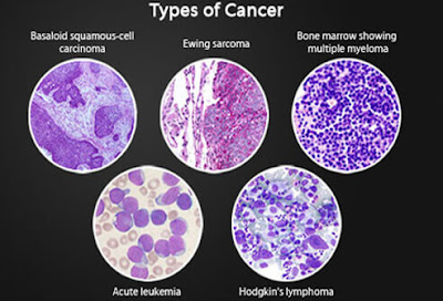
6 Types of Cancer

Cancer can occur anywhere in the body. Broadly, cancers are classified as either solid (for example breast, lung, or prostate cancers) or liquid (blood cancers). Cancer is further classified according to the tissue in which it arises.


1. Carcinoma

Carcinomas are cancers that occur in epithelial tissues in the body. They comprise 80% to 90% of all cancers. Most breast, lung, colon, skin, and prostate cancers are carcinomas. This class includes the two most common skin cancers, basal cell carcinoma and squamous cell carcinoma. Also in this class is the glandular cancer adenocarcinoma.


2. Sarcoma Cancer

Sarcomas occur in connective tissue like the bones, cartilage, fat, blood vessels, and muscles. This class of cancers includes the bone cancers osteosarcoma and Ewing sarcoma, Kaposi sarcoma (which causes skin lesions), and the muscle cancers rhabdomyosarcoma and leiomyosarcoma.


3. Myeloma Cancer

Myelomas are cancers that occur in plasma cells in the bone marrow. This class of cancer includes multiple myeloma, also known as Kahler disease.


 4. Leukemia

Leukemias are a group of different blood cancers of the bone marrow. They cause large numbers of abnormal blood cells to enter the bloodstream.


5. Lymphoma Cancer

Lymphomas are cancers of the immune system cells. These include the rare but serious Hodgkin lymphoma (Hodgkin’s lymphoma, also Hodgkin’s disease) and a large group of white blood cell cancers known collectively as non-Hodgkin lymphoma (non-Hodgkin’s lymphoma).


6. Mixed Cancer

Mixed cancers arise from more than one type of tissue.


How Stages of Cancer Are Determined


Doctors use the stages of cancer to classify cancer according to its size, location, and extent of spread. Staging helps doctors determine the prognosis and treatment for cancer. The TNM staging system classifies cancers according to:
  • Tumor (T): Primary tumor size and/or extent
  • Nodes (N): Spread of cancer to lymph nodes in the regional area of the primary tumor
  • Metastasis (M): Spread of cancer to distant sites away from the primary tumor
Some cancers, including those of the brain, spinal cord, bone marrow (lymphoma), blood (leukemia), and female reproductive system, do not receive a TNM classification. Instead, these cancers are classified according to a different staging systems.


What Are The Stages of Cancer? 


The TNM classification of a cancer usually correlates to one of the following five stages.
  • Stage 0: This refers to cancer that is "in situ," meaning that cancerous cells are confined to their site of origin. This type of cancer has not spread and is not invading other tissues.
  • Stage I – Stage III: These higher stages of cancer correspond to larger tumors and/or greater extent of disease. Cancers in these stages may have spread beyond the site of origin to invade regional lymph nodes, tissues, or organs.
  • Stage IV: This type of cancer has spread to distant lymph nodes, tissues, or organs in the body far away from the site of origin.


Diagnosing Cancer

Various tests may be performed in order to confirm a cancer diagnosis. Positron Emission Tomography and Computed Tomography (PET-CT) Scans and other similar tests can

highlight “hot spots” of cancer cells with high metabolic rates.
The most common test and procedures used to diagnose cancer include:
  • Mammogram
  • Pap Test
  • Tumor Marker Test
  • Bone Scan
  • MRI
  • Tissue Biopsy
  • PET-CT Scan

The Role of Lymph Nodes in Cancer Diagnosis

Cancer that originates in the lymph nodes or other area of the lymphatic system is called lymphoma. Cancer that originates elsewhere in the body can spread to lymph nodes. The presence of metastasized cancer in the lymph nodes is may mean the cancer is growing quickly and/or is more likely to spread to other sites. The presence of cancer in lymph nodes often affects prognosis and treatment decisions. Many diagnostic tests look at the lymph nodes as an indicator.


What Are Treatment Options?

The treatment is highly variable depending on the type and stage of a cancer as well as the overall health of the patient. The most common treatments are surgery, radiation, and chemotherapy. Other treatments include targeted/biological therapies, hematopoietic stem cell transplants, angiogenesis inhibitors, cryosurgery, and photodynamic therapy.
Every treatment has potential risks, benefits, and side effects. The patient and his or her care team, which may include an internist or other specialist, surgeon, oncologist, radiation oncologist, and others, will help determine the best and most appropriate course of treatment.
Until a cure can be found, prevention through a healthy lifestyle is the best way to stop cancer. Some ways to help protect yourself from cancer include eating plenty of fruits and vegetables, maintaining a healthy weight, abstaining from tobacco, drinking only in moderation, exercising, avoiding sun damage, getting immunizations, and getting regular health screenings.

  • Surgery

Surgery is often performed to remove malignant tumors. Surgery allows for the determination of the exact size of the tumor as well as the extent of spread and invasion into other nearby structures or lymph nodes – all-important factors in prognosis and treatment. Surgery is often combined with other cancer treatments, such as chemotherapy and/or radiation.
Sometimes, cancer cannot be entirely surgically removed because doing so would damage critical organs or tissues. In this case, debulking surgery is performed to remove as much of the tumor as is safely possible. Similarly, palliative surgery is performed in the cases of advanced cancer to reduce the effects (for example, pain or discomfort) of a cancerous tumor. Debulking and palliative surgeries are not curative, but they seek to minimize the effects of the cancer.
Reconstructive surgery can be performed to restore the look or function of part of the body after cancer surgery. Breast reconstruction after a mastectomy is an example of this kind of surgery.

  • Radiation Therapy

Radiation is a very common cancer treatment. About 50% of all cancer patients will receive radiation treatment, which may be delivered before, during, or after surgery
and/or chemotherapy. Radiation can be delivered externally -- where X-rays, gamma rays, or other high-energy particles are delivered to the affected area from outside the body -- or it can be delivered internally. Internal radiation therapy involves the placement of radioactive material inside the body near cancer cells. This is called brachytherapy.
Systemic radiation involves the administration of radioactive medication by mouth or intravenously. The radioactive material travels directly to the cancerous tissue. Radioactive iodine (I-131 for thyroid cancer) and strontium-89 (for bone cancer) are two examples of systemic radiation treatments.
Typically, external radiation is delivered 5 days a week over the course of 5 to 8 weeks. Other treatment regimens are sometimes used.

  • Chemotherapy Procedure

Chemotherapy, or "chemo," refers to more than 100 different medications used to treat cancer and other conditions. If eliminating all cancer cells is not possible, the goals of treatment may be to slow the growth of the cancer, keep the cancer from spreading,
and/or relieve cancer-associated symptoms (such as pain).
Depending on the type of chemotherapy prescribed, the medications may be given by mouth, injection, intravenously (IV), or topically. IV chemotherapy may be delivered via a catheter or port, which is usually implanted in a blood vessel of the chest for the duration of the therapy. Sometimes chemotherapy is delivered regionally, directly to the area that needs treatment. For example, intravesical therapy is used to infuse chemotherapy directly into the bladder for the treatment of bladder cancer.
The chemotherapy regimen a patient receives depends upon the type and stage of the cancer, any prior cancer treatment, and the overall health of the patient. Chemotherapy is usually administered in cycles over the course of days, weeks, or months, with rest periods in between.

Other Treatments

In addition to surgery, radiation, and chemotherapy, other therapies are used to treat cancer. These include:

  • Targeted or Biological Therapies

Targeted or biological therapies seek to treat cancer and boost the body's immune system while minimizing damage to normal, healthy cells. Monoclonal antibodies, immunomodulating drugs, vaccines, and cytokines are examples of targeted or biological therapies.

  • Hematopoietic Stem Cell Transplants

Hematopoietic stem cell transplants involve the infusion of stem cells into a cancer patient after the bone marrow has been destroyed by high-dose chemo and/or radiation.

  • Angiogenesis Inhibitors

Angiogenesis inhibitors are medications that inhibit the growth of new blood vessels that cancerous tumors need in order to grow.

  • Cryosurgery

Cryosurgery involves the application of extreme cold to kill precancerous and cancerous cells.

  • Photodynamic Therapy

Photodynamic therapy (PDT) involves the application of laser energy of a specific wavelength to tissue that has been treated with a photosensitizing agent, a medication that makes cancerous tissue susceptible to destruction with laser treatment. Photodynamic therapy selectively destroys cancer cells while minimizing the damage to normal, healthy tissues nearby.


Ongoing Research

Ongoing cancer research continues to identify newer, less toxic, and more effective cancer treatments. Visit the National Cancer Institute (NCI) to see a list of ongoing clinical trials.


 PET-CT (Positron Emission Tomography and Computed Tomography) Scan  :


Bone Metastasis


Bone metastasis happens in people who have cancer that forms in another part of the body. Cancer cells from that area break away and travel through the bloodstream and lymphatic system. When the cancer spreads to distant body organs, this is called metastasis. Common sites of metastasis include bones, the lungs, and the liver.


Risk Factors

Any type of cancer can raise the risk for bone metastasis. Although your doctor can't tell for sure if cancer will spread, certain types of cancer are more likely to spread to bone. These include cancers of the breast, lungs, thyroid, prostate, and kidneys. Larger tumors that have spread to the lymph nodes are also more likely to spread to the bones.


Where Bone Metastases Form

A bone metastasis can grow in any bone. But cancer most commonly spreads to the bones that are close to the center of the body. The spine is the most common place for a bone metastasis to form. Other common areas for bone metastases include the thigh bone, upper arm bone, ribs, hips, and skull.


Symptoms

Bone pain is often the first symptom of a bone metastasis. At first, the pain may come and go. It is often worse at night but feels better when you move. Over time, the pain may feel worse with movement and become more constant.


Cord Compression

Cancer growing in the spine can put pressure on the spinal cord. This can affect the nerves, causing loss of mobility, muscle weakness, numbness, and trouble urinating. Cord compression is a medical emergency because it can lead to paralysis. Treatments include steroid injections, radiation, chemotherapy, and surgery. Physical therapy may be needed to help regain muscle tone.


How Metastasis Affects the Bones

Bone metastasis often damages bones by making them weaker. Over time, this can cause them to break, even during routine activities like coughing or sitting down in a chair. At other times, bone metastasis can cause bone to become harder, though it may still break. In some cases, bone metastases can have both types of features.


Imaging Tests

Your doctor may order imaging tests to see if the cancer has spread to the bones, even if you haven't noticed any symptoms. These tests look at the inside of the body. X-rays may help find areas of bone metastases and confirm if a bone is broken. A bone scan can often find metastases earlier than an X-ray, and can check the whole body at once. CT scans, MRI scans, and PET scans also can show cancer that has spread.


Lab Tests

As bone metastases damage bone, calcium is often released from the bone into the bloodstream. High calcium levels can be a serious problem and can lead to nausea, constipation, dehydration, and even coma. Your doctor may send a tissue sample from the bone to a lab to confirm bone metastases.


Treating the Primary Cancer

Chemotherapy and hormone therapy target the main cancer. Taken by mouth or injection, they attack any cancer cells in the body. These treatments, called systemic because they affect the whole body, can cause side effects such as nausea, vomiting, fatigue, hair loss, and increased risk of infection.


Bisphosphonates

Bisphosphonates are a class of drugs that treats weak bones. They can be used to treat osteoporosis, a condition in which the bones get thin and weak. In certain cancer patients, bisphosphonates may help curb bone pain, reduce bone damage, lower elevated calcium levels, and decrease the risk for broken bones. Side effects include fatigue, nausea, vomiting, anemia, and bone or joint pain. A serious but rare side effect is jawbone death.


Treating Just the Bones

Local treatments focus on just the bone or bones where the cancer has spread. They work by destroying the tumor or slowing down the growth of the cancer cells. External beam radiation uses high-powered energy rays to kill cancer cells. It is similar to an X-ray, but much stronger. Ablation is another local treatment that destroys tumors with cold, heat, electric currents, or alcohol. Ultrasound may also be used to destroy nerve endings in the bone around the tumor. Another local treatment involves injecting bone cement to help stabilize the area.


Surgery

Surgery is often used as a treatment to help stabilize weakened bone that may be at risk for breakage. Your doctor may insert rods, screws, pins, or plates to help stabilize a bone and keep it from breaking. Surgery is also used to repair broken bones. But broken bones due to cancer often don't heal well. Your doctor may focus on trying to prevent bones from breaking in the first place.


Pain Management

In many cases, treatments for bone metastasis will also help relieve bone pain. But if pain persists, your doctor may suggest over-the-counter pain relievers, such as acetaminophen, ibuprofen, or naproxen. If these medications don't bring relief, your doctor may prescribe a stronger pain reliever, such as codeine, hydrocodone, oxycodone, or morphine.


After Treatment

Bone metastases aren't usually curable, but treatments may be able to shrink them and relieve symptoms. Talk to your health care provider about any new symptoms or side effects you've noticed. You may have a quicker recovery if you take an active role in your health -- by asking questions, learning more about your condition and its treatments, and taking care of yourself. Talk to your doctor about other treatment options that may be right for you.

01 Januari, 2017

Skin Cancer


Common Skin Cancer Symptoms and Causes
Sunlight contains ultraviolet light that is harmful to human skin cells. These energetic light waves can produce mutations in the DNA of skin cells, which in turn can lead to skin cancer. In areas close to the equator, the incidence of cutaneous cancers is dramatically higher due to the increase in sun exposure.
The most obvious skin cancer warning sign is the development of a persistent bump or spot in an area of sun-damaged skin. These spots are likely to bleed with minimal trauma and produce a superficial erosion.

Ultraviolet Light and Skin Cancer
Ultraviolet rays are classified by three types: UVA, UVB, and UVC. UVC is very dangerous, but it does not reach the earth’s surface due to the ozone layer. Exposure to both UVA and UVB radiation poses potential skin cancer risks.

UVA Radiation
UVA light is the most abundant source of solar radiation. Scientists think it can penetrate the top layer of skin, potentially damaging connective tissue and causing skin cancer. An estimated 50% of UVA exposure occurs in the shade. Light skin is far more vulnerable to UVA radiation: while dark skin allows only 17.5% of UVA to penetrate, light skin allows 55% of UVA light to pass through.

UVB Radiation
Sunburns are mostly caused by UVB radiation. Because of the ozone layer, UVB light accounts for only about 5% of the light that reaches the earth’s surface. UVB light can be filtered out by glass windows and does not penetrate as far into the skin as UVA, but it can still cause some forms of skin cancer. UVB is absorbed directly by DNA. Dark skin is twice as effective as light skin at protecting against UVB penetration.

How Skin Cancer Develops
UV light causes skin cancer by damaging the skin’s cellular DNA. That damage is caused by free radicals, which are hyperactive molecules found in UV light. Free radicals cause damage to the DNA double helix, changing the way cells replicate and naturally die, which is how cancer develops. In addition to sun exposure, free radicals are also found in environmental pollutants, cigarette smoke, alcohol, and other toxins. 

·        Actinic Keratosis (Solar Keratosis)

Actinic keratoses are scaling, horny, red, tender bumps present in sun-exposed areas. They are essentially very thin skin cancers that have not yet penetrated into deeper tissues. Large areas of skin thus exposed over years can result in what has recently been called a cancerization field defect. Continued exposure to ultraviolet light will likely induce invasive malignancies. Prevention and treatment of this condition which is most common in less-pigmented people is a significant part of medical skin care.

·        Actinic Cheilitis (Farmer's Lip)

Actinic cheilitis is simply actinic keratoses affecting the mucous membranes of the lips (vermillion border). This condition usually involves the lower lip simply because of the angle of incidence at which the overhead light waves hit the face. The forehead, cheeks, nose, and lower lip receive light waves perpendicularly and are not shaded by anatomical structures like the brows.

·        Cutaneous Horns

A cutaneous horn is a mass of dead skin cells. Essentially they have a lot in common with hair and nails since these are also composed of dead skin cells. The base which generates the horn can be an actinic keratosis, a squamous cell carcinoma, or a benign keratosis. The only way to differentiate between the three is by performing a surgical procedure called a biopsy and having it examined in a laboratory by a pathologist.

Identifying Cancerous Moles

The word "mole" is probably derived from a German word meaning spot. In the context of skin, a mole is a long-lasting skin spot that is a little darker than a freckle. Since there are a variety of benign and malignant skin growths which fulfill this description, more precision is necessary. The number of moles present on a particular person is most closely related to skin color and the extent of exposure to sunlight during childhood. The average number of moles is about 35 per person in Northern European populations.

·         Melanocytic Nevus
Moles that are melanocytic nevi (nevus singular) are local growths of melanocytes which can be present at birth or may develop after birth well into the third decade. They vary in color from black to flesh-colored. A melanocytic nevus can develop into melanoma, particularly if the nevus is large. The risk of a congenital melanocytic nevus developing into melanoma is greater for children under 10, which account for 70% of all cases.

·        Atypical Moles

When a physician examines a particular spot or mole it can appear either ordinary or peculiar. Most of these atypical nevi -- when examined under the microscope -- are not cancer; it seems to be the new lesions that develop in adulthood that are the ones that are most likely to be malignant melanomas.
The term dysplastic refers to changes noted in moles that can only be appreciated on microscopic examination. This term should only be used when describing the microscopic appearance of a nevus or other tissue. Most atypical nevi exhibit some degree of dysplasia under the microscope.

ABCDEs of Melanoma

In an attempt to systemize nevus description physicians use a variety of adjectives to describe visible alterations. To simplify things, benign lesions are almost always uniform in color, circular in shape, and exhibit bilateral symmetry about any axis drawn through its diameter. Malignant melanomas diverge from this appearance to a greater or lesser extent. To help one remember the criteria, they have been called the ABCDEs (mnemonic) of nevus description. Another valuable consideration is the so-called "ugly duckling" nevus which appears significantly different than all the other skin spots on the patient.

Melanoma Symptoms


>> 'A' Is for Asymmetry

Asymmetry refers to the degree of similarity when one compares the appearance of each of four quadrants produced by an imaginary cross through the middle of a melanocytic nevus.


>> 'B' Is for Border

Border refers to degree of circular regularity of the edge of a pigmented lesion. Perfectly circular lesions are rarely malignant.


>> 'C' Is for Color

Color: The degree of uniformity of color is a measure of atypicality. The more colors present the more likely the lesion is to be malignant.


>> 'D' Is for Diameter

Diameter: Although malignant pigmented lesions tend to be larger than a pencil eraser this is one of the less reliable criteria for distinguishing melanomas. 


>> 'E' Is for Evolving

Evolving: Malignancies by their very nature tend to change over time -- mostly by increasing in size -- as opposed to benign lesions that are stable. Since cancers grow in an uncontrolled way they tend to produce asymmetrical lesions.


Screening for Cancerous Moles

If a mole looks or acts at all peculiarly it is best to have it evaluated by an expert. This frequently is a dermatologist. Most dermatologists can tell if the pigmented lesion is composed of melanocytes or is something quite different with no possibility of being a melanoma. Many dermatologists now use a hand-held magnifying device which produces polarized light to evaluate colored melanocytic tumors. The use of this instrument improves the doctor's ability to identify suspicious lesions.

Evaluating Potentially Cancerous Moles

The decision on which mole needs to be examined depends on the concern the doctor has that it may be cancerous. Since the procedure necessary to take off the suspicious bump is simple, requiring only local anesthesia and rarely is associated with complications, the threshold for this procedure is appropriately low. If the lesion is melanocytic in origin and it appears at all peculiar, exhibiting any of the features addressed in the ABCDE criteria and/or the patient complains that the bump bleeds or is irritated or itchy, that is often enough to trigger a biopsy.

Malignant Melanoma

Malignant melanoma is the most deadly form of skin cancer because it tends to spread at a very early stage to other parts of the body. The likelihood that it has spread is most directly related to the thickness of the melanoma as measured from its surface to its depth by a pathologist. This information is communicated to the doctor that submitted the nevus and subsequent treatment depends on this information.


Melanoma Symptoms
In addition to the ABCDEs of melanoma, there are other melanoma symptoms that may include:
  • A sore that will not heal
  • Pigment that spreads from the border of a spot into the surrounding skin
  • Redness or swelling around a skin spot
  • A spot that becomes itchy, tender, or painful
  • Scaliness, bleeding, or oozing from the surface of a mole


How Melanoma Cancer Develops

Although it is possible for a previously benign mole to become malignant, most of the time a melanoma cancer begins as a single malignant cell in previously normal skin and then continues to grow in an uncontrolled manner.


Squamous Cell Carcinoma

Squamous cell carcinomas begin as malignancies of living epidermal cells which grow and invade into the deeper layers of the skin. They usually begin as an actinic keratosis and progress over many years. Although most actinic keratoses do not eventuate into squamous cell cancers, if one has a great many of them and they continue to receive sufficient ultraviolet irradiation producing sufficient mutational events, it is extremely likely that a squamous cell carcinoma will develop. They appear as thick, keratotic bumps on sun-exposed skin which continue to enlarge. They do not commonly spread to distant sites, but larger lesions can do this.

Bowen Disease: Is it Cancer?

So called "Bowen's disease" lesions are simply squamous cell carcinomas that have not as yet penetrated into the deeper layers of the skin. They involve the entire thickness of the most superficial living layer of the epidermis as opposed to actinic keratoses which involve the upper half of the epidermis. They seem to be a stage in the progression of an actinic keratosis to an invasive squamous cell carcinoma. As such it is cancer. Bowen disease is easily curable by cryotherapy, curettage with cautery and other methods.

Basal Cell Carcinoma

Basal cell carcinomas arise from the layer of skin cells present along the lowest layer of the epidermis and are the most common form of skin cancer. They are locally aggressive so they ought to be treated before they get so large that removal becomes difficult. The tumor has a shiny surface, is pearly white in color, and tends to bleed quite easily. It often ulcerates.


Who Is At Risk for Skin Cancer and Why?

Sunlight is by far the most common cause of skin cancer. Most of the exposure occurs during leisure time or sun tanning. The perceived benefits of exposure to sunlight seem to be confined mostly to the production of vitamin D in the skin and a belief that darker skin is more aesthetically appealing. Vitamin D requirements can be easily satisfied with dietary supplementation. Light-skinned individuals with blonde or red hair who live in regions near the equator are the most at risk to develop skin cancer.

Reduce Your Skin Cancer Risk
Beyond living a healthy lifestyle, it is important to avoid exposure to sunlight as much as is reasonably possible. Eating a balanced diet and staying physically active improves the body’s ability to fight free radical damage and heal.

Protecting Your Skin
Any time there has been a darkening of the skin after sun exposure it is a sign that some damage has been sustained. Since the incubation period for skin cancers and photo-aging is quite long (5 to 15 years), it may be difficult to convince sun worshipers to head indoors.

Avoiding UV Damage
The use of clothing, searching for shade, and the application of effective, broad-spectrum sunscreens are all useful behaviors in limiting sun damage.

Seeking Shade to Avoid Skin Cancer
Shade is the first defense against skin damage. Between the hours of 10 a.m. and 4 p.m., when the sun’s rays are at their most intense, find cover or wear a wide-brimmed hat. The Skin Cancer Foundation recommends hats with broad brims all around that are at least three inches wide.
Although shade is an important protection against skin cancer, it may still leave you vulnerable to UVB light, which can reach skin indirectly. UV radiation can bounce off of clouds, dry sand, concrete and other UV-reflective surfaces.

Choosing the Right Sunscreen
Sunscreens that are reasonably durable are currently available that block all the wavelengths of ultraviolet light with an SPF (Sun Protection Factor) of 50. It can help to look for the words “broad spectrum” or “multi spectrum” when choosing a sunscreen. This indicates that both UVA light and UVB light are being blocked by your sunscreen. Keep in mind that these phrases don’t actually indicated how much of each ray is being blocked. Also, consider using an FDA approved “Water Resistant (40 or 80 min.)” sunscreen if you sweat or swim in sunlight.