31 Desember, 2016

Ovarian Cancer


This cancer begins in the ovaries, the twin organs that produce a woman's eggs and the main source of the female hormones estrogen and progesterone. Treatments for ovarian cancer have become more effective in recent years, with the best results seen when the disease is found early.

Ovarian Cancer Symptoms

  • Bloating or pressure in the belly.
  • Pain in the abdomen or pelvis.
  • Feeling full too quickly during meals.
  • Urinating more frequently.
These symptoms can be caused by many conditions that are not cancer. If they occur daily for more than a few weeks, report them to your health care professional.

Risk Factor:

~ Family History

A woman's odds of developing ovarian cancer are higher if a close relative has had cancer of the ovaries, breast, or colon. Researchers believe that inherited genetic changes account for 10% of ovarian cancers. This includes the BRCA1 and BRCA2 gene mutations, which are linked to breast cancer. Women with a strong family history should talk with a doctor to see whether closer medical follow-up could be helpful.

~ Age

The strongest risk factor for ovarian cancer is age. It's most likely to develop after a woman goes through menopause. Using postmenopausal hormone therapy may increase the risk. The link seems strongest in women who take estrogen without progesterone for at least 5 to 10 years. Doctors are not certain whether taking a combination of estrogen and progesterone boosts the risk as well.

~ Obesity

Obese women have a higher risk of getting ovarian cancer than other women. And the death rates for ovarian cancer are higher for obese women too, compared with non-obese women. The heaviest women appear to have the greatest risk.

Ovarian Cancer Screening Tests

There are two ways to screen for ovarian cancer before it causes symptoms or shows up during a routine gynecologic exam. One is a blood test for elevated levels of a protein called CA-125. The other is an ultrasound of the ovaries. Unfortunately, neither technique has been shown to save lives when used in women of average risk. For this reason, screening is only recommended for women with strong risk factors.

Diagnosing Ovarian Cancer

Imaging tests, such as ultrasound or CT scans (seen here), can help reveal an ovarian mass. But these scans can't determine whether the abnormality is cancer. If cancer is suspected, the next step is usually surgery to remove suspicious tissues. A sample is then sent to the lab for further examination. This is called a biopsy. Sometimes a sample taken with a needle can also be used for diagnosis.

Stages of Ovarian Cancer

The initial surgery for ovarian cancer also helps determine how far the cancer has spread, described by the following stages:
  • Stage I : Confined to one or both ovaries.
  • Stage II : Spread to the uterus or other nearby organs.
  • Stage III : Spread to the lymph nodes or abdominal lining.
  • Stage IV : Spread to distant organs, such as the lungs or liver.
Types of Ovarian Cancer
The vast majority of ovarian cancers are epithelial ovarian carcinomas. These are malignant tumors that form from cells on the surface of the ovary. Some epithelial tumors are not clearly cancerous. These are known as tumors of low malignant potential (LMP.) LMP tumors grow more slowly and are less dangerous than other forms of ovarian cancer.

Ovarian Cancer Survival Rates

Ovarian cancer can be a frightening diagnosis, with 5-year relative survival rates that range from 89% to 18% for epithelial ovarian cancer, depending on the stage when the cancer was found. But keep in mind that these odds are based on women diagnosed from 1988 to 2001. The treatments and outlook may be better for people diagnosed today. For LMP tumors, the five-year relative survival rates range from 99% to 77%.

Ovarian Cancer Surgery

Surgery is used to diagnose ovarian cancer and determine its stage, but it is also the first phase of treatment. The goal is to remove as much of the cancer as possible. This may include a single ovary and nearby tissue in stage I. In more advanced stages, it may be necessary to remove both ovaries, along with the uterus and surrounding tissues.

Chemotherapy

In all stages of ovarian cancer, chemotherapy is usually given after surgery. This phase of treatment uses drugs to target and kill any remaining cancer in the body. The drugs may be given by mouth, through an IV, or directly into the belly (intraperitoneal chemotherapy.) Women with LMP tumors usually don't need chemo unless the tumors grow back after surgery.

Targeted Therapies

Researchers are working on therapies that target the way ovarian cancer grows. A process called angiogenesis involves the formation of new blood vessels to feed tumors. A drug called Avastin blocks this process, causing tumors to shrink or stop growing (seen in the illustration here). Avastin is approved for other cancers, but ovarian cancer researchers are still testing this therapy, which can have serious side effects.

After Treatment:

~ Early Menopause

When women have both ovaries removed, they can no longer produce their own estrogen. This triggers menopause, no matter how young the patient. The drop in hormone levels can also raise the risk for certain medical conditions, including osteoporosis. It's vital that women have regular follow-up care after being treated for ovarian cancer.

~ Moving On

Women may find that it takes a long time for their energy to return after treatments end. Fatigue is a very common problem after treatment for cancer. Beginning a gentle exercise program is one of the most effective ways to restore energy and improve emotional well-being. Check with your health care team to determine which activities are right for you.

Risk Reducer:

~ Pregnancy

Women who have biological children are less likely to get ovarian cancer than women who have never given birth. The risk appears to decrease with every pregnancy, and breastfeeding may offer added protection.

~ 'The Pill'

Ovarian cancer is also less common in women who have taken birth control pills. Women who have used the pill for at least five years have about half the risk of women who never took the pill. Like pregnancy, birth control pills prevent ovulation. Some researchers think ovulating less often may protect against ovarian cancer.

~ Tubal Ligation

Getting your tubes tied, formally known as tubal ligation, may offer some protection against ovarian cancer. The same goes for having a hysterectomy – removing the uterus while leaving the ovaries intact.

~ Removing the Ovaries

For women with genetic mutations that put them at high risk for ovarian cancer, removing the ovaries is an option. This can also be considered in women over 40 getting a hysterectomy.

~ Low-Fat Diet

While there is no definitive diet to prevent ovarian cancer, there is evidence that what you eat can make a difference. In one recent study, women who stuck to a low-fat diet for at least four years were less likely to develop ovarian cancer. Some researchers report the cancer is also less common in women who eat a lot of vegetables, but more studies are needed.


Breast Cancer


Breast Cancer Symptoms

There are often no symptoms of breast cancer, but sometimes women may discover a breast problem on their own. Signs and symptoms to be aware of may include:
  • A painless lump in the breast.
  • Changes in breast size or shape.
  • Swelling in the armpit.
  • Nipple changes or discharge.
Breast pain can also be a symptom of cancer, but this is not common.

Signs of Inflammatory Breast Cancer

Inflammatory breast cancer is a rare, fast-growing type of cancer that often causes no distinct lump. Instead, breast skin may become thick, red, and may look pitted -- like an orange peel. The area may also feel warm or tender and have small bumps that look like a rash.


Breast Cancer and Mammogram

The earlier breast cancer is found, the easier it is to treat. And mammogram, X-rays of the breast, can detect tumors before they are large enough to feel. The American Cancer Society recommends yearly mammogram beginning at age 40 for women at average risk. While the U.S. Preventive Services Task Force recommends a screening mammogram every two years from age 50 to 74. It also notes that before age 50, each woman should check with a doctor to find out what screening schedule is right for her, considering the potential benefits and harms from screening.


Breast Ultrasound and MRI

Besides a mammogram, your doctor may order additional imaging with breast ultrasound. An ultrasound can help determine the presence of cysts, fluid-filled sacs that are not cancer. An MRI may be recommended along with a mammogram for routine screening in certain women who have a higher risk of breast cancer.


Breast Self-Exams

It was once widely recommended that women check their own breasts once a month. But studies suggest these breast self-exams play a very small role in finding cancer. The current thinking is that it’s more important to know your breasts and be aware of any changes, rather than checking them on a regular schedule. If you want to do breast self-exams, be sure to go over the technique with your doctor.


What If You Find a Lump?

First, don’t panic. Eighty percent of breast lumps are not cancerous. Lumps often turn out to be harmless cysts or tissue changes related to your menstrual cycle. But you should let your doctor know right away if you find anything unusual in your breast. If it is cancer, the earlier it’s found the better. And if it’s not, testing can give you peace of mind.


Breast Biopsy

The only sure way to determine whether a lump is cancer is to do a biopsy. This involves taking a tissue sample for further examination in the lab, sometimes through a small needle. Sometimes surgery is done to take part of or the entire lump for testing. The results will show whether the lump is cancer, and if so, what type. There are several forms of breast cancer, and treatments are carefully matched to the type of cancer.


Hormone-Sensitive Breast Cancer

Some types of breast cancer are fueled by the hormones estrogen or progesterone. A biopsy can reveal whether a tumor has receptors for estrogen (ER-positive) and/or progesterone (PR-positive). About two out of three breast cancers are hormone sensitive. There are several medications that keep the hormones from promoting further cancer growth.
The image shows a molecular model of an estrogen receptor.


HER2-Positive Breast Cancer

In about 20% of patients, breast cancer cells have too many receptors for a protein called HER2. This type of cancer is known as HER2-positive, and it tends to spread faster than other forms of breast cancer. It’s important to determine whether a tumor is HER2-positive, because there are special treatments for this form of cancer.


Breast Cancer Stages

Once breast cancer has been diagnosed, the next step is to determine how big the tumor is and how far the cancer has spread. This process is called staging. Doctors use Stages 0-4 to describe whether cancer is localized to the breast, has invaded nearby lymph nodes, or has spread to other organs, such as the lungs. Knowing the stage and type of breast cancer will help your health care team formulate a treatment strategy.


Breast Cancer Survival Rates

The odds of surviving breast cancer are strongly tied to how early it is found. According to the American Cancer Society, 100% of women with Stage 1 breast cancer live at least five years, compared to women without cancer – and many women in this group remain cancer-free for good. The more advanced the cancer, the lower this figure becomes. By Stage 4, the five-year relative survival rate declines to 20%. But these rates can improve as more effective treatments are found.


Breast Cancer Surgery

There are many types of breast cancer surgery, from taking out the area around the lump (lumpectomy or breast-conservation surgery) to removing the entire breast (mastectomy.) It’s best to discuss the pros and cons of each of these procedures with your doctor before deciding what’s right for you.


Radiation Therapy 

Radiation therapy uses high-energy rays to kill cancer cells. It may be used after breast cancer surgery to wipe out any cancer cells that remain. It can also be used along with chemotherapy for treatment of cancer that has spread to other parts of the body. Side effects can include fatigue and swelling or a sunburn-like feeling in the treated area.


Chemotherapy 

Chemotherapy uses drugs to kill cancer cells anywhere in the body. The drugs are often given by IV, but are sometimes taken by mouth or shot. Chemotherapy may be done after surgery to lower the odds of the cancer coming back. In women with advanced breast cancer, chemotherapy can help control the cancer’s growth. Side effects may include hair loss, nausea, fatigue, and a higher risk of infection.


Hormone Therapy

Hormone therapy is an effective treatment for women with ER-positive or PR-positive breast cancer. These are cancers that grow more rapidly in response to the hormones estrogen or progesterone. Hormone therapy can block this effect. It is most often used after breast cancer surgery to help keep the cancer from coming back. It may also be used to reduce the chance of breast cancer developing in women who are at high risk.


Targeted Drugs

Targeted therapies are newer drugs that target specific properties within cancer cells. For example, women with HER2-positive breast cancer have too much of a protein called HER2. Targeted therapies can stop this protein from promoting the growth of cancer cells. These drugs are often used in combination with chemotherapy. They tend to have milder side effects compared to chemotherapy.


Life After Diagnosis

There’s no doubt that cancer is a life-changing experience. The treatments can wear you out. You may have trouble managing daily chores, work, or social outings. This can lead to feelings of isolation. It’s crucial to reach out to friends and family for support. They may be able to go with you to treatments, help out with chores, or just remind you that you are not alone. Many people choose to join a support group -- either locally or online.


Breast Reconstruction

Many women who have a breast removed choose to undergo reconstructive surgery. This replaces the skin, nipple, and breast tissue that are lost during a mastectomy. Reconstruction can be done with a breast implant or with tissue from somewhere else in your body, such as the tummy. Some women opt to begin reconstruction at the same time as their mastectomy. But it’s also possible to have reconstructive surgery months or years later.

Breast Forms

An alternative to breast reconstruction is to be fitted for a breast form. This is a breast-shaped prosthesis that fits inside your bra. Wearing a breast form allows you to have a balanced look when you are dressed -- without undergoing additional surgery. Like reconstructive surgery, breast forms are often covered by insurance.


Breast Cancer: Why Me?

The most obvious risk factor for breast cancer is being a woman. Men get the disease, too, but it is about 100 times more common in women. Other top risk factors include being over age 55 or having a close relative who has had the disease. But keep in mind that up to 80% of women with breast cancer have no family history of the illness.

Breast Cancer Genes

Some women have a very high risk of breast cancer because they inherited changes in certain genes. The genes most commonly involved in breast cancer are known as BRCA1 and BRCA2. Women with mutations in these genes have up to an 80 percent chance of getting breast cancer at some point in life. Other genes may be linked to breast cancer risk as well.

Risk Factors in Your Control

Being overweight, getting too little exercise, and drinking more than one alcoholic beverage per day can raise the risk of developing breast cancer. Birth control pills and some forms of postmenopausal hormone therapy can also boost your risk. But the risk goes back to normal after these medications are stopped. Among survivors, good lifestyle choices may be helpful. Recent studies suggest that physical activity may help lower the risk of a recurrence and it's a proven mood-booster.

Breast Cancer Research

Doctors continue to search for more effective and tolerable treatments for breast cancer. The funding for this research comes from many sources, including advocacy groups throughout the country. Many of the 2.5 million breast cancer survivors and their families choose to participate in walk-a-thons and other fundraising events. This links each individual fight against cancer into a common effort for progress.


Note :


Hormonal Therapy

If your cancer is fueled by hormones such as estrogen, you may need to take hormonal therapy. You and your doctor might choose from various types of these drugs. They can lower the amount of estrogen your body makes so it's not available to fuel your breast cancer.
There are different types of hormonal treatments for breast cancer:


Tamoxifen and toremifene (Fareston) block estrogen from stimulating cancer cell growth. Doctors call these drugs "SERMS," which stands for selective estrogen receptor modulators.


Anastrozole (Arimidex), exemestane (Aromasin), and letrozole (Femara) stop the body from making estrogen in women who've gone through menopause.


Fulvestrant (Faslodex) counters estrogen throughout the body, not just in cancer cells. You get it in a shot. It's approved for use in postmenopausal women with advanced breast cancer who've already tried tamoxifen or toremifene.


Goserelin (Zoladex) and leuprolide (Lupron) stop the ovaries from making estrogen. Doctors may consider these meds, along with other hormone drugs, in women who have not yet been through menopause. After stopping these drugs, the ovaries may or may not make estrogen again.


Targeted Treatments

There are drugs that target specific proteins related to cancer.


Everolimus (Afinitor) targets a protein called mTOR, and palbociclib (Ibrance) goes after a protein called CDK 4/6. They're approved to treat advanced breast cancer in women after menopause if:

  • Their disease is sensitive to (meaning fueled by) estrogen. Doctors call this “ER-positive.” Most breast cancers are ER-positive.
  • Their cancer is not sensitive to the HER2 protein. Your doctor may call this “HER2-negative.” Most breast cancers are HER2-negative.


Some breast cancers -- about 20% -- make too much of the HER2 protein. They're more aggressive than other cancers. Drugs that target HER2 include:
 
Trastuzumab (Herceptin) blocks the HER2 protein from stimulating cancers cells to grow. You get it by IV once a week or every 3 weeks as a bigger dose. One of the risks is congestive heart failure, so your doctor will closely watch your heart's health if you take it.

Ado-trastuzumab emtansine (TDM-1, Kadcyla) is like trastuzumab with a chemo drug added to it. You get it by IV every 3 weeks.

Pertuzumab (Perjeta) works similarly to trastuzumab by blocking HER2. Doctors often give it along with the chemo drug docetaxel (Taxotere) and trastuzumab.
 
Lapatinib (Tykerb) may be an option if chemo and trastuzumab aren't working.


Other Treatments

If the disease is in your bones, you may need another medication, such as:

Denosumab (Prolia, Xgeva). This drug will slow down the growth of breast cancer in your bones, and it protects bones from breaking. It can also lower the blood's calcium level, so your doctor will monitor this. You get a shot of it, usually every 4 weeks.

Pamidronate disodium (Aredia). When breast cancer is in the bones, you may have too much calcium in your blood. This drug lowers your blood's calcium level. You get it by IV, usually every 3-4 weeks. Each session can take 2 or more hours, depending on your particular case.

Zoledronic acid (Zometa). This is the same type of drug as pamidronate disodium. It works the same way to lower your blood calcium level. You get it by IV, which takes about 15 minutes, every 3-4 weeks.
Talk with your doctor about the risks and benefits of each treatment. Keep your personal goals in mind as you decide how best to treat your cancer.


Breast Cancer Surgery (animation) :


Breast Reconstruction Surgery Following Mastectomy


29 Desember, 2016

Bladder Cancer


Bladder cancer is the growth of abnormal or cancerous cells on the inner lining of the bladder wall. Most bladder cancers are detected at early stages when the tumor has not spread outside the bladder and treatments are successful.


Symptoms: 


Blood in Urine (Hematuria)
One sign of bladder cancer is blood in the urine, also known as hematuria. Blood in the urine does not always mean bladder cancer. Hematuria is most often caused by other conditions like trauma, infection, blood disorders, kidney problems, exercise, or certain medications. Blood in the urine may be seen by the naked eye (gross hematuria) or only detected on urine testing (microscopic hematuria). The urine may be discolored and appear brownish or darker than usual or, rarely, bright red in color.


Bladder Changes

Bladder cancer sometimes causes changes in bladder habits like having to urinate more often or feeling an urgent need to urinate without producing urine. Another symptom of bladder cancer is pain or burning during urination without evidence of a urinary tract infection. These symptoms of bladder problems, like bleeding, are usually caused by conditions other than cancer. Bladder cancer tends to cause no symptoms until it reaches an advanced stage that is difficult to cure.


 Possible Causes of Bladder Cancer:

·        Smoking

Smoking is the greatest known risk factor for bladder cancer; smokers are four times more likely to get bladder cancer than nonsmokers. Harmful chemicals from cigarette smoke enter the bloodstream in the lungs and are ultimately filtered by the kidneys into the urine. This leads to a concentration of harmful chemicals inside the bladder. Experts believe that smoking causes about half of all bladder cancers in men and women.

·        Chemical Exposure

Exposure to certain chemicals on the job can increase risk of bladder cancer. Occupations that may involve exposure to cancer-causing chemicals include metal workers, hairdressers, and mechanics. Organic chemicals called aromatic amines are especially associated with bladder cancer and are used in the dye industry. Those working with dyes, metal workers, or in the manufacturing of leather, textiles, rubber, or paint should be sure to follow recommended safety protocols. Smoking increases the risk even more for these workers.


Who is at Risk for Bladder Cancer?

Bladder cancer can affect anyone, but certain groups are at greater risk. Men are three times more likely than women to get bladder cancer. Around 90% of cases occur in people over age 55, and whites are twice as likely as African Americans to develop the condition.
Other factors that increase the risk of getting bladder cancer include a family history of the condition and previous cancer treatment. Birth defects involving the bladder increase the risk of bladder cancer. When people are born with a visible or invisible defect that connects their bladder with another organ in the abdomen, this leaves the bladder prone to frequent infection. This increases the bladder’s susceptibility to cellular abnormalities that can lead to cancer. Chronic bladder inflammation (frequent bladder infections, bladder stones, and other urinary tract problems that irritate the bladder) increase the risk of developing bladder cancer.


Diagnosis:

>> Testing

There is no single lab test that can specifically screen for and diagnose bladder cancer, even though urine tests may suggest that cancer is present. If a cancer is present, several tests may be abnormal, including urine cytology and tests for tumor marker proteins.


  • Cystoscopy

A type of endoscopy, cystoscopy, is a procedure that allows visualization of the inside of the bladder through a thin, lighted tube that contains a camera. The instrument can also take small samples (biopsies) if abnormal areas are seen. A tissue biopsy is the most reliable way to diagnose bladder cancer.


  • Urinalysis and Urine Cytology

An analysis of the urine is a very useful test in the diagnosis of and screening for many diseases and conditions. The urinalysis will detect any abnormalities in the urine such as blood, protein, and sugar (glucose). A urine cytology is the examination of urine under a microscope while looking for abnormal cells that might indicate bladder cancer.

>> Imaging

  • Intravenous Pyelogram

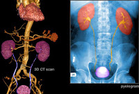
An intravenous pyelogram is an X-ray test with contrast material (dye) to show the uterus, kidneys, and bladder. When testing for bladder cancer, the dye highlights the organs of the urinary tract allowing physicians to spot potential cancer-specific abnormalities.
  • CT Scans and MRI

CT scans and MRI are often used to identify tumors and trace metastasized cancers as they spread to other organ systems. A CT scan provides a three-dimensional view of the bladder, the rest of the urinary tract, and the pelvis to look for masses and other abnormalities. CT scans are often used in conjunction with Positron emission tomography (PET)to highlight cells with high metabolic rates. “Hot spots” of cells with abnormally high metabolism may indicate the presence of cancer and require further investigation.
  • Bone Scan

If a tumor is found in the bladder a bone scan may be performed to determine whether the cancer has spread to the bones. A bone scan involves having a small dose of a radioactive substance injected into the veins. A full body scan will show any areas where the cancer may have affected the skeletal system.


Types of Bladder Cancer

Bladder cancers are named for the specific type of cell that becomes cancerous. Most bladder cancers are transitional cell carcinomas, named for the cells that line the bladder. Other less common types of bladder cancer are squamous cell carcinoma and adenocarcinoma.

  • Transitional Cell Carcinoma

Bladder cancer that begins inside the innermost tissue layer of the bladder, the transitional epithelium, is known as transitional cell carcinoma. This type of lining cell is are able to stretch when the bladder is full and shrink when it is emptied. Most bladder cancers begin in the transitional epithelium.
There are two types of transitional cell carcinoma, low-grade and high-grade. Low-grade transitional cell carcinoma tends to come back after treatment, but rarely spreads into the muscle layer of the bladder or to other parts of the body. High-grade transitional cell carcinoma also tends to come back after treatment and will often spread into the muscle layer of the bladder, other parts of the body, and the lymph nodes. High-grade diseases cause most bladder cancer deaths.
  • Squamous Cell Carcinoma

Squamous cells are thin, flat cells that may lead to bladder cancer after irritation or long-term infection.
  • Adenocarcinoma

Adenocarcinoma cancers emerge from glandular cells in the lining of the bladder. Adenocarcinoma is a very rare form of bladder cancer.


Stages of Bladder Cancer 


Cancer staging is typically determined by the extent to which a cancer has grown or spread. A staging system is a way for professionals to specifically describe how much a cancer has progressed. Typically, the TNM system is used for bladder cancer and represents the following:
  • T describes how far the main tumor has grown
  • N reveals any cancer spread to lymph nodes near the bladder
  • M reveals whether the cancer has spread (metastasized) to other locations away from the bladder.


Bladder Cancer Stages

Stage 0a (Ta, N0, M0): The cancer is non-invasive papillary carcinoma and has not invaded the connective tissue or bladder wall muscle.
Stage 0is (Tis, N0, M0): Cancerous cells in the inner lining tissue of the bladder only.

Stage I (T1, N0, M0): Tumor has spread onto the bladder wall.

Stage II (T2, N0, M0): Tumor has penetrated the inner wall and is present in muscle of the bladder wall.

Stage III (T3, N0, M0): Tumor has spread through the bladder to fat around the bladder.

Stage IV applies to one of the following: (T4, N0, M0): Tumor has grown through the bladder wall and into the pelvic or abdominal wall.

Any T, N1, M0: The tumor has spread to the nearby lymph nodes. Any T, any N, M1: The tumor has spread to distant lymph nodes or to sites such as bones, liver, or lungs.


Bladder Cancer Treatment:


>>  Surgery

  • Transurethral Resection

Early stage cancers are most commonly treated by transurethral surgery. An instrument (resectoscope) with a small wire loop is inserted through the urethra and into the bladder. The loop removes a tumor by cutting or burning it with electrical current, allowing it to be extracted from the bladder.
  • Partial and Radical Cystectomy

Partial cystectomy includes the removal of part of the bladder. This operation is usually for low-grade tumors that have invaded the bladder wall but are limited to a small area of the bladder. In a radical cystectomy, the entire bladder is removed, as well as its surrounding lymph nodes and other areas that contain cancerous cells. If the cancer has metastasized outside of the bladder and into neighboring tissue, other organs may also be removed such as the uterus and ovaries in women and the prostate in men.


>>  Urinary Diversion After Surgery

When the entire bladder is removed the surgeon will create an alternate way for urine to be stored and passed. This procedure is called urinary diversion. Depending on preference, a bag can either be placed inside or outside of the body to collect urine. Non-continent urinary diversion is when a urostomy bag is placed outside the body, worn under the clothes. Continent urinary diversion consists of a pouch, made from intestinal tissue, inside the body to hold urine. In a newly introduced surgical procedure, the insertion of an artificial bladder has also been successful for some patients.


>> Chemotherapy

Chemotherapy is given in some cases before surgery to shrink bladder cancer tumors. It can also be used after surgery to destroy any remaining tumor cells. Chemotherapy may be given intravenously or administered directly into the bladder (intravesical chemotherapy). Intravesical chemotherapy is effective in decreasing the recurrence rate of superficial bladder cancers on a short-term basis, but not effective against bladder cancer that has already invaded the muscular walls. Systemic or intravenous chemotherapy is required when the cancer has deeply penetrated the bladder, lymph nodes, or other organs.

 Chemotherapy Side Effects

Side effects vary from patient to patient. Common side effects of systemic chemotherapy include the following:
  • Nausea andvomiting
  • Loss of appetite
  • Hair loss
  • Sores on the inside of the mouth or in the digestive tract
  • Feeling tired or lacking energy
  • Increased susceptibility to infection
  • Easy bruising or bleeding
  • Numbness or tingling in the hands orfeet


>>  Immunotherapy

Immunotherapy involves the administration of helpful bacteria through a catheter into the bladder to trigger the immune system to attack both the bacteria and the cancer cells. Immunotherapy is only given in stages Ta, T1, and CIS (carcinoma in situ) bladder cancers. Bacillus Calmette-Guerin (BCG) is a type of bacteria used in this therapy. Intravesical BCG treatment is given once a week and can be used after surgery to lower the chance of tumor recurrence. Immunotherapy side effects can include irritation of the bladder, minor bleeding in the bladder, and flu-like symptoms.

·      

 >>  Radiation

What is Radiation?

Radiation therapy is the use of painless, invisible, high-energy radiation that can kill both healthy and cancerous cells. Radiation can be used as an alternative approach or in addition to chemotherapy or surgery to destroy cancer cells. 

  • External Radiation

External radiation is produced by a machine outside the body. The machine aims a concentrated beam of radiation at the tumor. External radiation is typically given five days a week for five to seven weeks. 

  • Internal Radiation

Internal radiation consists of inserting a small pellet of radioactive material inside the bladder. The treatment lasts several days and patients are required to stay in the hospital until the pellet is removed. 

Radiation Side Effects

Radiation therapy also has side effects, which can include fatigue, nausea, skin irritation, pain with urination, and diarrhea.

Alternative Treatments for Bladder Cancer

There are no alternative or complementary therapies that have been shown to prevent or cure bladder cancer. Ongoing research studies are examining the role of green tea or broccoli sprouts as potential complementary treatments. 

Bladder Cancer Survival Rates and Prognosis

Bladder Cancer Survival Rates

As with most cancers, survival rates are dependent upon the stage or extent of spread of the cancer when it is found. About 50% of bladder cancers are detected when the tumor is limited to the inner lining of the bladder, and 5-year survival rates for this early stage of cancer are nearly 100%. Cancers that have spread further typically have lower survival rates. Today the relative survival rates for all stages of bladder cancer are 77% at 5 years, 70% at 10 years, and 65% at 15 years.

Bladder Cancer Prognosis

The outlook for bladder cancer patients depends on the stage of cancer at the time of diagnosis. Patients with metastatic bladder cancer that has spread to other organs have an average life expectancy of 12 to 18 months. Recurrent cancer suggests a more aggressive type and a negative outlook for long-term survival for patients with advanced stage bladder cancer.


Sex After Bladder Cancer Treatment

The surgery for bladder cancer can damage nerves in the pelvis, making sex difficult.

Changes for Men

Some men may have trouble getting an erection, but in younger men, this may improve over time. Semen cannot be produced if the surgery involved removal of the prostate gland and seminal vesicles.

Changes for Women

In women, the uterus, ovaries, and part of the vagina are removed during radical cystectomy. This permanently stopsmenstruation and prohibits all future pregnancies. Women who undergo surgery for bladder cancer may also find that sex is less comfortable, and achieving orgasms may be difficult.


Bladder Cancer Prevention

There is no known way to prevent bladder cancer, but it is always advisable to follow a healthy lifestyle. Stop smoking and limit alcohol consumption to 1 to 2 drinks a day. A healthy diet contains lots of fruits, vegetables, whole grains, and correct portion sizes of lean meats. Regular exercise and having checkups can also help you support your health and provide peace of mind. Avoid unsafe chemical exposures and keep protected if working with chemicals.


New and Experimental Treatments for Bladder Cancer

New treatments are being investigated for bladder cancer. These include photodynamic therapy, gene therapy, and targeted therapy. Clinical trials are available to test some of these or other new therapies.
  • Photodynamic Therapy

Photodynamic therapy uses a laser light and chemicals to kills cancer cells and shrink tumors. A few days before treatment, the patient is given light-sensitive compounds intravenously that sensitizes cancer cells to the light rays emitted by a laser. A small scope with a laser is then introduced into the bladder through the urethra and is aimed at the tumor.
  • Gene Therapy

Gene therapy refers to the introduction of cells with laboratory-altered DNA into the body in order to prevent the mutation and spread of cancerous cells or to attack cancerous cells and tumors by cutting off blood supply or causing internal cellular death to targeted cancer cells. Gene therapy often requires the use of a patient’s blood or bone marrow may in order to perform the procedure. Experimental in nature, gene therapy is a newly emerging procedure with a growing research base. Some scientists believe gene therapy may be the best way to find a cure for cancer.
  • Targeted Therapy

Targeted therapies are directed at limiting growth of cancer cells. Targeted therapy uses drugs to interfere with specific molecules involved in carcinogenesis and tumor growth.